Équipes de recherche et développement associés au RISEst

La R&D et le RISEst

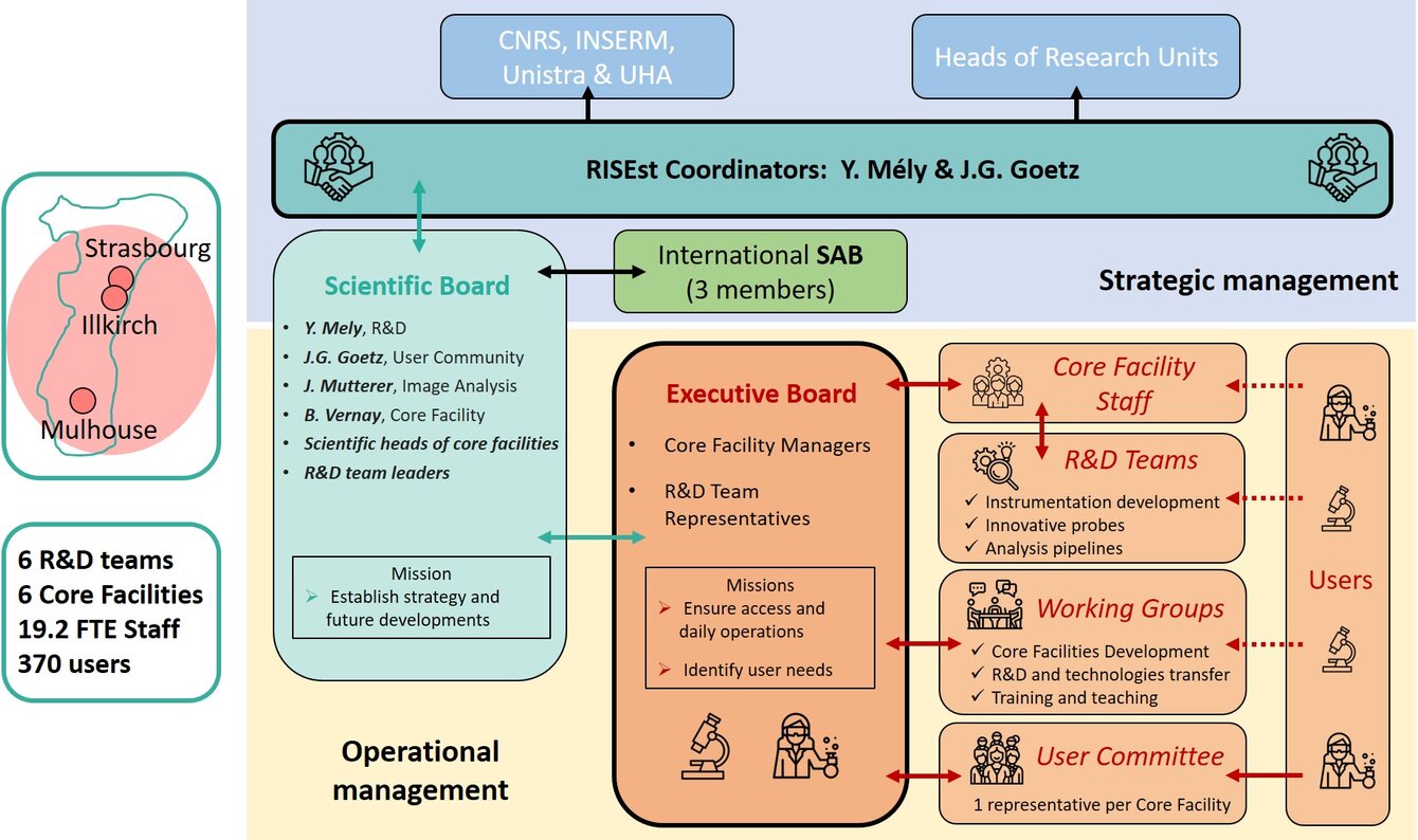
Six équipes de recherche et développement de renommée interationale sont en relation étroite avec le RISEst. Elles développent des technologies et des méthodologies innovantes et assurent leur transfert vers les plateformes.

Équipes de développement technologiques

IMTI : Imagerie Microscopique et Traitement d'Images (Lien)

Laboratoire : IRIMAS Institut de Recherche en Informatique, Mathématiques, Automatique et Signal
Contact : olivier.haeberle[at]uha.fr
Adresse : IRIMAS – Université de Haute-Alsace  12 rue des Frères Lumière  68 093 MULHOUSE Cedex

Projet

The IMTI team (IRIMAS) has developed tomographic diffractive microscopy techniques for imaging unlabelled samples. Such systems achieve 3D refractive index distribution measurements at cellular level, with twice-improved resolution compared to conventional optical microscopes. IMTI accomplished several world-first achievements in correlative TDM-Fluorescence microscopy, high lateral resolution/high longitudinal precision profilometry, isotropic resolution TDM images (≈180 nm in 3D), distinguishing absorption and refraction and experimental mirror-assisted TDM images.

Biophotonique des interactions moléculaires et cellulaires (lien)

Laboratoire : LBP Laboratoire de Biophotonique et Pathologies
Contact : yves.mely[at]unistra.fr
Adresse : LBP Faculté de Pharmacie 74 route du Rhin - CS 60024    F-67401 Illkirch-Graffenstaden cedex

Projet

The Biophotonics team (LBP) has developed anti-Stokes wide-field and luminescence lifetime confocal microscopes to track and image upconverting nanoparticles. The team also recently implemented a spectral PAINT super-resolution (SR) microscopy, a light sheet single molecule microscope and a single molecule UV microscope. Together, with the previously developed two-photon FLIM/FCS and 3D PALM/STORM set-ups, this allowed the team to provide the Quest facility with a state-of-the-art set of quantitative and SR microscopes. Moreover, LBP is currently implementing a PAINT methodology with high-speed acquisition and low background noise using DRET (Dark Resonance Energy Transfer).

IPP Photonics Instrumentation and Processes (Lien)

Laboratoire : iCUbe laboratoire des sciences de l'Ingénieur, de l'Informatique et de l'Imagerie
Contact : sylvain.lecler[at]insa-strasbourg.fr  / maioli[at]unistra.fr
Adresse :  Télécom Physique Strasbourg Pôle API   300, Boulevard Sébastien Brant  67412 Illkirch

Projet

The Photonics Instrumentation and Processes team (IPP-ICube), has a long-standing expertise in microscopy and coherent imaging, as well as diffuse optics for surgical guidance. IPP has notably developed full-field OCT (scanning interference microscopy), endoscopic OCT, digital holography, hyperspectral imaging and quantitative phase imaging for biomedical applications. The team has a particular interest in multimodal imaging and contrast, which includes spectroscopy, elastography and Doppler imaging. The team has also more recently started working on fluorescence imaging, and notably light-sheet microscopy for high-throughput imaging of live biomedical samples, based on the single-objective Oblique Plane Microscopy technique.

Large complexes involved in transcription and translation team (Lien)

Laboratoire : IGBMC Institut de Génétique et Biologie Moléculaire et Cellulaire
Contact : klaholz[at]igbmc.fr
Adresse :  LBP - UMR 7021 Faculté de pharmacie, 74 route du Rhin  67401 Illkirch-Graffenstaden Télécom Physique Strasbourg Pôle API   300, Boulevard Sébastien Brant  67412 Illkirch

Projet

The Large complexes involved in transcription and translation team (IGBMC) has recently performed a series of developments in SR fluorescence microscopy to facilitate single molecule localization microscopy (SMLM) analysis with the aim of integrating structural data generated through crystallography, cryo-EM, cryo-ET and FIB towards the cellular level. This team notably developed an integrated software for image reconstruction, drift and chromatic aberration correction, co-localization, resolution estimation (SharpViSu), segmentation and clustering of labelled complexes [ClusterViSu], including 3D analysis and segmentation of SMLM data using Voronoi diagrams. In addition, the team is currently developing a spectral demixing method, which facilitates co-localization of proteins in SMLM.

Équipes de développement méthodologiques

Nanochemistry and Bioimaging (Lien)

Laboratoire : LBP Laboratoire de Biophotonique et Pathologies
Contact : andrey.klymchenko[at]unistra.fr
Adresse : LBP Faculté de Pharmacie 74 route du Rhin - CS 60024    F-67401 Illkirch-Graffenstaden cedex

Projet

The Nanochemistry and Bioimaging team (LBP) has notably developed innovative solvatochromic probes for ratiometric and super-resolution imaging as well as fluorogenic probes for advanced fluorescence microscopy techniques. More specifically, the team designed, synthesized and characterized a large set of polarity-sensitive membrane probes based on different molecular scaffolds for sensing lipid organization in plasma membranes, polarity mapping, monitoring cell stress (oxidative and starvation) and polarity sensing in organelles (ER, Golgi, Lipid Droplets, mitochondria, lysosomes). The team also designed a family of bright probes for plasma membranes and extracellular vesicles compatible with classical and super-resolution imaging techniques, bright fluorescent markers of lipid droplets, red calcium probes, fluorogenic probes for G-protein coupled receptors and RNA aptamers (o-Coral). In parallel, this team has developed dye-loaded fluorescent polymeric nanoparticles (NPs), much brighter than quantum dots, for Single Particle Tracking in live cells and mice brain, barcoding of living cells and intracellular RNA imaging. Finally, the team developed fluorescent nanoemulsion droplets of different colours for in vivo imaging.

Synpa  Synthesis for analysis (Lien)

Laboratoire : IPHC Institut pluridisciplinaire Hubert Curien
Contact : l.charbonn[at]unistra.fr
Adresse : LBP Faculté de Pharmacie 74 route du Rhin - CS 60024    F-67401 Illkirch-Graffenstaden cedex

Projet

The Synpa team (IPHC) is developing luminescent labels for microscopy imaging based on lanthanides and thus, characterized by very long-lived excited states, line-like emission spectra and large pseudo Stokes’ shift. The team especially focuses on the design of luminescent probes displaying upconversion at the molecular and supramolecular level as well as on the optimization of lanthanide-based nanoparticles for time-resolved luminescence microscopy. The molecular approach has already provided exciting molecular sized constructs devoid of the problems associated to the use of nanoparticles (biotoxicity, reproducibility,…) while benefiting of the spectroscopic advantages of the anti-Stokes process.

These R&D teams in probe development are already actively interacting with the facilities and other R&D teams of the node, in order to validate their tools and test them in biological applications. For instance, there is a close collaboration between the Biophotonics and the SynPa teams for the development of molecular upconverting complexes and their characterization and imaging by the developed anti-Stokes microscopes transferred to the Quest facility. Moreover, a large number of the probes developed by the Nanophotonics team have been validated on the high resolution and quantitative microscopes developed by the Biophotonics teams and transferred to different users of the imaging facilities both in Illkirch and Strasbourg.